
AMD is a common eye condition among people age 50 and older. It is a leading cause of vision loss in older adults. It gradually destroys the macula, the part of the eye that provides sharp, central vision needed for seeing objects clearly.
In some people, AMD advances so slowly that vision loss does not occur for a long time. In others, the disorder progresses faster and may lead to a loss of vision in one or both eyes. The vision loss makes it difficult to recognize faces, drive a car, read, print, or do close work, such as sewing or fixing things around the house.
Despite the limited vision, AMD does not cause complete blindness. You will be able to see using your side (peripheral) vision.
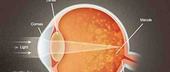
The Macula
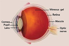
The macula is made up of millions of light-sensing cells that provide sharp, detailed central vision. It is the most sensitive part of the retina, which is located at the back of the eye. The retina quickly turns light into electrical signals and then sends these electrical signals to the brain through the optic nerve. Next, the brain translates the electrical signals into images we see. If the macula is damaged, fine points in these images are not clear. The picture is there, but the fine points are lost.
Who is at risk?
AMD usually occurs in people who are age 50 and older. As people get older, the risk increases. Other risk factors include the following:
-
•Smoking. Research shows that smoking increases the risk of AMD two-fold.
-
•Race. Caucasians are much more likely to get AMD than people of African descent.
-
•Family history. People with a family history of AMD are at higher risk.
Does lifestyle make a difference?
Some lifestyle choices, like smoking, are linked to AMD although it remains unknown if altering any of these would alter the impact of AMD on an individual. Nevertheless, the following choices may have an impact on AMD and certainly promote healthy living, including the following:
-
•Avoiding smoking
-
•Exercising
-
•Maintaining normal blood pressure and cholesterol levels
-
•Eating a healthy diet rich in green, leafy vegetables and fish
How is AMD detected?
The early and intermediate stages of AMD usually start without symptoms. Only a comprehensive dilated eye exam can detect AMD. The eye exam may include the following:
-
•Visual acuity test. This eye chart measures how well you see at distances.
-
•Dilated eye exam. Your eye care professional places drops in your eyes to widen or dilate the pupils. This gives him or her a better view of the back of your eye. Using a special magnifying lens, he or she then looks at your retina and optic nerve for signs of AMD and other eye problems.
-
•Amsler grid. Your eye care professional also may ask you to look at an Amsler grid. Changes in your central vision may cause the lines in the grid to disappear or appear wavy, a sign of AMD.
-
•OCT
-
•Fluorescein angiogram. Your eye care professional may suggest you see an ophthalmologist to perform a fluorescein angiogram. With this test, your doctor injects a dye into your arm. Pictures are taken as the dye passes through the blood vessels in your eye. The test allows your doctor to identify leaking blood vessels and decide the best treatment.
What are the forms of AMD that can cause vision loss?
There are two forms of AMD: dry and wet. Either form can advance and cause severe vision loss. Later sections of this booklet describe the different types in greater detail.
The following is a brief description of each:
-
•The dry form is more common and has three stages-early, intermediate, and advanced. It happens when the light-sensitive cells in the macula slowly break down, gradually blurring central vision in the affected eye.
-
•The wet form is considered advanced AMD and can be more severe. It happens when new blood vessels under the macula leak blood and fluid. Damage to the macula can occur rapidly.
All people who have the wet form had the dry form first.
Dry AMD
What is dry AMD?
Dry AMD is the most common form of AMD in its early or intermediate stages. It occurs in about 90 percent of the people with the condition.
Dry AMD happens when the light-sensitive cells in the macula slowly break down, gradually blurring central vision in the affected eye. As dry AMD progresses, you may see a blurred spot in the center of your vision. Your eye care professional may call this “geographic atrophy.”
Over time, central vision in the affected eye can be slowly lost as less of the macula works.
What are the symptoms?
Dry AMD has few symptoms in the early stages. It is important to have your eyes examined regularly before the disease progresses.
In the later stages, blurred vision is the most common symptom of dry AMD. Objects also may not appear to be as bright as they used to be.
As a result, you may have trouble recognizing faces. You may need more light for reading and doing other tasks. Both eyes can have dry AMD or one eye can be affected first.
What are drusen?
Drusen are another early sign of dry AMD. They are yellow deposits under the retina. They can be small or large in size.
Your eye care professional can see drusen when he or she examines the retina during a comprehensive dilated eye exam.
Drusen alone do not usually cause vision loss. But people with large drusen are at risk of developing a more severe form of AMD, which results in severe vision loss.
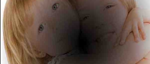
Normal vision

The same scene as viewed by a person with age-related macular degeneration
Three stages of dry AMD
Dry AMD has three stages, all occurring in one or both eyes.
These stages are defined in part by the size and number of drusen under the retina:
-
•Early AMD. People with early AMD have either small drusen or a few medium-sized drusen. At this stage, you may not have any symptoms or vision loss.
-
•Intermediate AMD. People with this stage of AMD have either many medium-sized drusen or one or more large drusen. Many people will have no symptoms, so don’t wait for symptoms to determine if you have an intermediate stage of AMD. Some people see a blurred spot in the center of their vision. They often need more light to read and to do other tasks.
-
•Advanced dry AMD. In addition to drusen, people with advanced dry AMD have a breakdown of light-sensitive cells supporting tissue in the macula. This breakdown can cause a blurred spot in the center of your vision, often called geographic atrophy. Over time, the blurred spot may get bigger and dark, taking away a larger area of your straight-ahead vision.
Vision loss and dry AMD
If you have vision loss from dry AMD in one eye only, you may not notice any changes in your overall vision. With the other eye seeing clearly, you still can drive, read, and see fine details.
You may notice changes in your vision if dry AMD affects both eyes or if you develop the wet form of the disease. In any case, see an eye care professional for a comprehensive dilated eye exam if blurring occurs in your vision.
Can the dry form turn into the wet form?
All people who have the wet form had the intermediate stage of the dry form first. The dry form also can suddenly turn into the wet form, even during early stage AMD. Eye care professionals have no way to tell if the dry form will turn into the more severe wet form.
Dry AMD can turn into wet AMD at any time. You should get an Amsler grid from your eye care professional to check your vision for signs of wet AMD.
Diet might help
Studies have shown that people who eat a diet rich in green, leafy vegetables and fish have a lower risk of developing AMD.
While there is no definitive proof that changing your diet will reduce your risk of developing AMD or having it progress, to maintain good health in general, there is no reason not to eat a healthy diet, exercise, avoid smoking, and see your healthcare professional regularly.
A note about early stage dry AMD
Currently, no treatment exists for early stage dry AMD, which in many people shows no symptoms or loss of vision. Your eye care professional may recommend that you get a comprehensive dilated eye exam at least once a year. The exam will help determine if your condition is advancing.
If your condition gets worse, your eye care professional may suggest that you take a specific high-dose supplement that contains antioxidants and zinc. Do not take these high-dose supplements unless your doctor recommends them. Research shows that high doses of specific vitamins and minerals may slow the condition’s progress
Wet AMD
What is wet AMD?
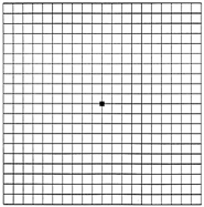
Here is what an Amsler grid normally looks like.
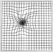
This is what an Amsler grid might look like to someone with AMD.
Wet AMD affects about 10 percent of all people with AMD. This type, however, is more severe than the early and intermediate stages of the dry form.
Wet AMD happens when abnormal blood vessels behind the retina start to grow under the macula. These new blood vessels can be fragile and leak blood and fluid. The blood and fluid cause the macula to swell and damage occurs rapidly. The damage may also cause scarring of the retina.
Although loss of central vision can happen quickly, eye care professionals can slow down or stop the progression of wet AMD if it is detected before severe vision loss occurs.
What are the symptoms?
During the early stages of wet AMD straight lines may appear wavy. People with wet AMD also may develop a blind spot, which results in the loss of central vision.
If you notice these or other changes to your vision, contact your eye care professional at once. Again, eye care professionals may be able to treat the condition before severe vision loss occurs.
Treatment options for wet AMD
With early diagnosis and proper treatment, you can delay the progression of AMD. The earlier it is detected, the better your chances of keeping your vision. Wet AMD typically results in severe vision loss. However, eye care professionals can try different therapies to stop further vision loss. You should remember that the therapies described below are not a cure. The condition may progress even with treatment.
-
•Injections. One option to slow the progression of wet AMD is to inject drugs into your eye. With wet AMD, abnormally high levels of vascular endothelial growth factor (VEGF) are secreted in your eyes. This substance promotes the growth of new abnormal blood vessels. The anti-VEGF injection therapy blocks its effects. If you get this treatment, you may need multiple injections. Your eye care professional may give them monthly. Before each injection, your eye care professional will numb your eye and clean it with antiseptics. To prevent the risk of infection, a doctor may prescribe antibiotic drops.
-
•Photodynamic therapy. This technique involves laser treatment of select areas of the retina. First, a drug called verteporfin will be injected into a vein in your arm. The drug travels through the blood vessels in your body, including any new, abnormal blood vessels in your eye. Your eye care professional then shines a laser beam into your eye to activate the drug in the blood vessels. Once activated, the drug destroys the new blood vessels and slows the rate of vision loss. This procedure takes about 20 minutes.
-
•Laser surgery. Eye care professionals sometimes treat certain cases of wet AMD with laser surgery, though this is less common than other treatments. This treatment is performed in a doctor’s office or eye clinic. It involves aiming an intense beam of light at the new blood vessels in your eyes to destroy them. However, laser treatment also may destroy some surrounding healthy tissue and cause more blurred vision.
Advanced AMD
What is advanced AMD?
Both the wet form and the advanced dry form are considered advanced AMD. It can occur in the same eye or an eye may have just one form or the other. In most cases, only advanced AMD can cause vision loss.
Additional risk
People who have advanced AMD in one eye are at especially high risk of developing advanced AMD in the other eye.
However, research has shown that high doses of vitamins and mineral supplements may slow the progression of intermediate AMD to the more advanced stage.
Age-Related Eye Disease Study
NEI research found that patients taking high doses of antioxidants and zinc could reduce their risk of developing advanced AMD and experiencing severe vision loss. The findings were based on a10-year clinical trial called the Age-Related Eye Disease Study (AREDS).
What is the AREDS formulation?
The AREDS formulation is a combination of antioxidants and zinc. The specific daily amounts of antioxidants and zinc tested in the AREDS clinical trial included the following:
-
•500 milligrams of vitamin C
-
•400 International Units of vitamin E
-
•15 milligrams of beta-carotene (often labeled as the equivalent of 25,000 International Units of vitamin A)
-
•80 milligrams of zinc as zinc oxide
-
•2 milligrams of copper as cupric oxide
This formulation is commonly taken with meals in the morning and evening in two equally divided doses.
Researchers added copper to the AREDS formulation to prevent copper-deficiency anemia. This condition happens in people who take high levels of zinc.
Taking multivitamins
Diet alone will not provide the same high levels of the vitamins and minerals found in the AREDS formulation. Multivitamins also do not contain the same high levels.
Multivitamins do contain many important vitamins not found in the AREDS formulation.
Therefore, you may want to take a multivitamin along with the AREDS formulation. If you are already taking daily multivitamins and your doctor suggests you take the high-dose AREDS formulation, be sure you review your vitamin supplements with your eye care professional before you begin.
Who should take the AREDS formulation?
The study showed that the formulation was most beneficial for people who had:
-
•Intermediate AMD in one or both eyes
-
•Advanced AMD (dry or wet) in one eye, but not the other eye
People with early stage AMD did not benefit from taking the AREDS formulation.
A note about the AREDS formulation
Researchers stress that the AREDS formulation is not a cure. It will not restore vision already lost from the condition. But it may delay the onset of advanced AMD. It also may help people who are at a high risk of developing advanced AMD keep their remaining vision.
Loss of Vision
Coping with AMD and vision loss can be a traumatic experience. This is especially true of those who have just begun to lose their vision or have low vision. Having low vision means that even with regular glasses, contact lenses, medicine, or surgery, people find everyday tasks difficult to do. Reading the mail, shopping, cooking, seeing the TV, and writing can all seem challenging.
However, help is available. You may not be able to restore your vision, but low vision services can help you make the most of what is remaining. You can continue enjoying friends, family, hobbies, and other interests just as you always have. The key is not delaying use of these services.
What is low vision rehabilitation?
Low vision rehabilitation is a specialized practice. It usually includes ophthalmologists and optometrists who received special training in low vision services. Specialists in low vision also work as a team with eye care professionals, social workers, vision rehabilitation teachers, and others to help people cope with failing eyesight (see box on The low vision team).
The first step in getting services is a low vision examination. It is different from normal eye exams. During this appointment, the specialist in low vision will do the following:
-
•Examine your medical history and ask questions about your daily routines, hobbies, and goals
-
•Thoroughly test your vision to make sure current prescriptions work
-
•Introduce you to low vision devices for seeing objects that are near and distant
-
•Make referrals for additional services, such as counseling and support groups
By testing your vision and asking questions, the team is better able to choose a low vision device that would work best for you. Training can take place at the specialist’s office, or a specialist will schedule a visit to set up and demonstrate equipment in your home. While there, he or she may evaluate lighting, mark appliances, and look for safety problems.
The low vision team
Specialist in Low Vision-An ophthalmologist or optometrist who is trained to evaluate and treat low vision.
Occupational Therapist-A licensed professional who teaches you how to use low vision devices.
Rehabilitation Teacher-An educator who teaches you different ways to handle the demands of daily living.
Orientation and Mobility (O&M) Professional-A professional who specializes in technologies that enable safe travel.
Social Worker-A professional who helps you cope with vision loss and emotional stress.
Technology Specialist or Adaptive Technology Specialist-A professional who is an expert in assistive and adaptive devices.
Where to go for services
Low vision services can take place in different locations, including the following:
-
•Ophthalmology or optometry offices that specialize in low vision
-
•Hospital clinics
-
•State, nonprofit, or for-profit vision rehabilitation organizations
-
•Independent-living centers
How to find a specialist
To find a specialist in low vision, talk with your eye care professional. He or she may be able to refer you to a variety of local resources to assist you.
What are some low vision devices?
Because low vision varies from person to person, specialists have different tools to help patients deal with vision loss.
They include the following:
-
•Reading glasses with high-powered lenses
-
•Handheld magnifiers
-
•Video magnifiers
-
•Computers with large-print and speech-output systems
-
•Large-print reading materials
-
•Talking watches, clocks, and calculators
Keep in mind that low vision aids without proper diagnosis, evaluation, and training may not work for you. It is important that you work closely with your low vision team to get the best device or combination of aids to help improve your ability to see.
Computer aids and other technologies
Today, computers are helping people with low vision with reading and writing. New technology can increase text size on computer screens and allow computers to read what appears on the screen. People with low vision also are benefiting from scan-to-read devices, note takers, and other advanced technologies.